
Table of contents
Today, an integral part of the work of a reproductive doctor is an ultrasound examination. Why is it so important and effective?
Is ultrasound safe?
Yes, the method of ultrasound diagnostics is absolutely safe and very informative. The essence of the method is the use of ultrasound, which comes from the probe, then the ultrasound waves, passing through the tissues to the organ, are reflected from it, and return back. The probe perceives them in the reverse direction and forms an image on the screen. Modern ultrasound devices use low-frequency waves that do not pose a threat.
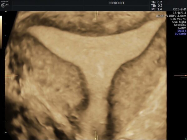
- T-shaped uterus (class U1a according to ESHRE)
Who should have an ultrasound of the pelvic organs?
To all women who are planning pregnancy. This non-invasive method of diagnosis is extremely informative, because with its help we can learn the following:
• Is the anatomical structure (structure) of the uterus correct
• What is the ovarian reserve of a woman (how many antral follicles are visualized in the ovaries), does it correspond to the reproductive age
• Is the endometrium normal (absence of polyps, hyperplasia, synechiae in the uterine cavity, presence of sufficient thickness for embryo implantation)
• Is there an inflammatory process in the fallopian tubes (please mention that fallopian tubes normally shouldn’t be visualized on ultrasound)
• Are there leiomyomas (nodes) in the uterus that can deform its cavity
• Are there any ovarian cysts (functional, endometrioid, dermoid, etc.)
• Is ovulation occurring (presence of a leading follicle or corpus luteum, depending on the phase of the menstrual cycle)
On which day of the menstrual cycle is it best to conduct an ultrasound examination?
Ideal for this method of diagnosis will be the first phase of the cycle (6-12), since we can best assess the endometrium to rule out or confirm polyps. However, the second phase (16-24 days of the menstrual cycle) will be useful in order to understand the thickness of the endometrium, whether it grows enough in the second phase of the cycle, or if it is too lush, thickened – then we are talking about endometrial hyperplasia. And the middle of the cycle (12-14-15 days) will be indicative in order to estimate on which day of the cycle ovulation occurs.
It is follicles that are visualized on ultrasound in any phase of the menstrual cycle. Therefore, the doctor can choose when it is best to conduct an ultrasound individually in your case.
If the ultrasound shows many follicles in the ovaries, is this PCOS?
No, multifollicular ovarian structure does not mean polycystic ovary syndrome (PCOS).
Additional criteria are required to establish this diagnosis:
• Laboratory signs of hyperandrogenism (increased level of male sex hormones in the blood)
• Clinical manifestations of hyperandrogenism (hirsutism, acne, alopecia)
• Oligo- or anovulation (extremely rare or no ovulations)
• The volume of the ovaries is more than 10 cm3
Ovarian cyst and persistence of non-ovulated follicle
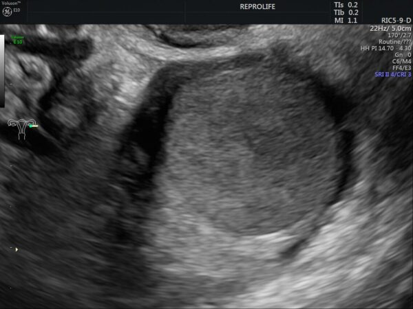
Ultrasound is the simplest and most effective method of differentiating between a functional (non-ovulating follicle) and a true cyst (endometrioid, dermoid ovarian cyst, teratoma, etc.). In the case of primary visualization of a cystic formation in the ovary, it will be advisable to conduct ultrasound in the next menstrual cycle for its final differentiation. A functional cyst does not require treatment, especially surgical removal. When conducting an ultrasound in the next menstrual cycle, it should decrease in size or disappear, but a real cyst will not have dynamics until the volume decreases.
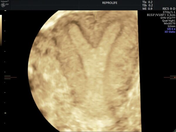
- Endometrioid ovarian heterotopia (endometrial ovarian cyst)
Is transabdominal ultrasound important in reproductive medicine?
With the invention of transvaginal ultrasound, it has become the method of choice in reproductive medicine. The anatomical structure of the uterus, endometrium, ovaries – all this is much better visualized precisely with transvaginal ultrasound, especially 3D. However, there are still situations when a transabdominal probe comes to the rescue: embryo transfer (embryo transfer is carried out with a full bladder), assistance is often needed ultrasound during hysteroscopic myomectomy (removal of uterine nodes that deform its cavity), pipel biopsy (examination of the endometrium in order to rule out the inflammatory process – endometritis), intrauterine insemination, atypical location of the ovaries, myomatous nodes that deform the uterine cavity.
- Endometrial polyp in 3D imaging
Can fallopian tubes be seen during ultrasound?
Unfortunately no. During ultrasound, fallopian tubes normally are not visualized, because they are very small (10-12 cm long), and the diameter of their lumen is only 2-6 millimeters. We can see them only if the lumen of the tube is expanded, filled with liquid (hydrosalpinx, pyosalpinx).

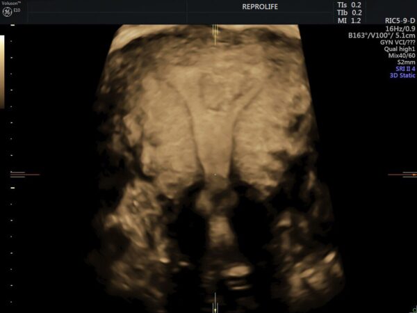
- 3D image of the uterine cavity with visualization of tubal angles
The main application of ultrasound in IVF programs
It should be noted that one of the most important stages of the IVF program is carried out with the help of an ultrasound machine. Follicle puncture (egg collection) is performed using a transvaginal probe with a special nozzle, and embryo transfer (embryo transfer into the uterine cavity) is performed under the control of a transabdominal probe.
It is very important to note that the level of performance and experience of the specialist who performs ultrasound is very important for the correct diagnosis and subsequent treatment tactics. Equally important is the level of ultrasound equipment. In our Medical Center, we work only on GE Voluson E 10 expert equipment, which provides the best quality of ultrasound diagnostics and the possibility of obtaining 3D images in gynecology of the highest possible quality available in medicine today for our patients.