Table of contents
Thanks to the timelapse system, we can monitor each stage of embryo development, starting from the first hours after fertilization and continuing until the blastocyst stage. This method allows for non-invasive observation of cell division and the dynamics of embryo development, assessing its potential for successful implantation.
Day One:
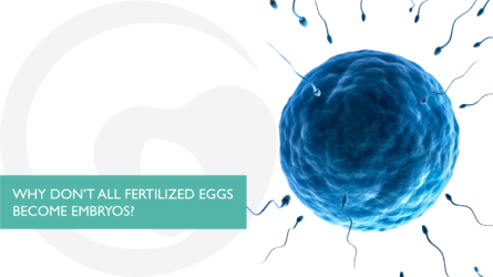
After fertilization, the egg and sperm fuse to form the first cell, the zygote, which contains genetic material from both parents.
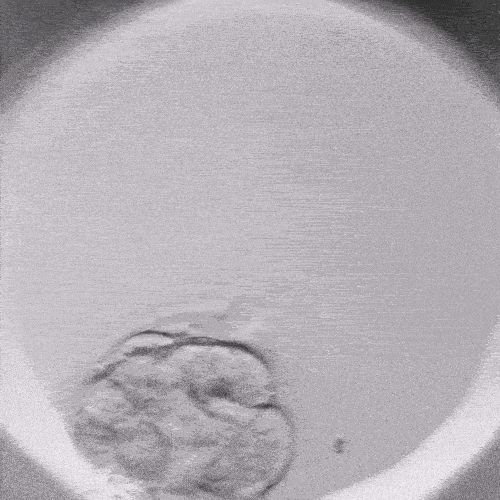
Day Two-Three:
The embryo undergoes cell division. The proper rhythm and uniformity of division are critically important.
Day Four:
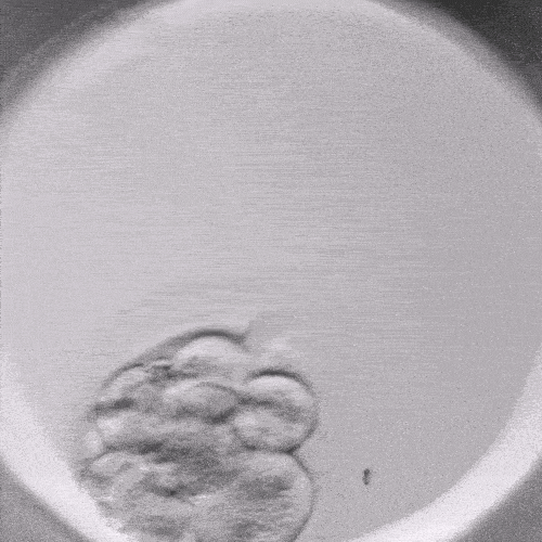
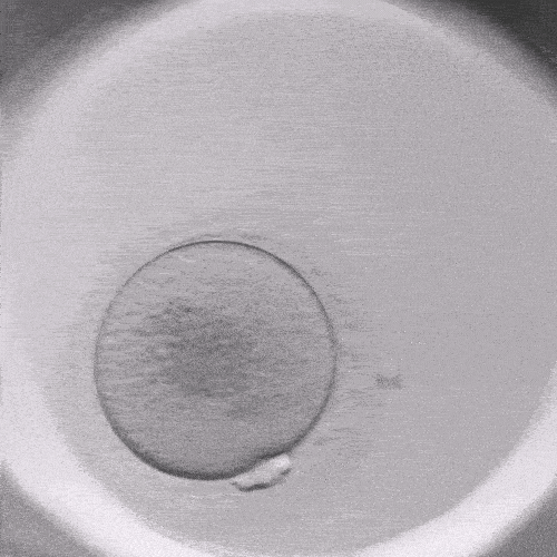
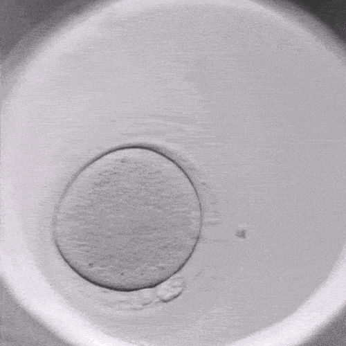
The embryo reaches the morula stage, where the cells merge to form a dense structure, making it difficult to distinguish individual cells.
Day Five:
The embryo reaches the blastocyst stage, where an inner cell mass and a cavity are formed. At this stage, the embryo is ready for transfer to the uterus or cryopreservation for future cycles.
What Are the Advantages of Timelapse Technology?
- Monitoring embryo development without removing them from the incubator minimizes stress and risks.
- Detailed information on morphology and development dynamics helps embryologists accurately assess embryo quality.
- The ability to select the best embryos for transfer increases the chances of a successful pregnancy.
- Visual data helps patients better understand the treatment process.
Each of these stages is crucial for assessing embryo quality. Timelapse technology allows doctors to observe development without harming the embryo and to select the best ones for transfer.