CHANGES IN THE STRUCTURE OF THE UTERUS – HOW THE UTERUS FORMS AND WHY SOMETIMES IT DOESN'T GO ACCORDING TO PLAN

Changes in the structure of the uterus – how the uterus forms and why sometimes it doesn’t go according to plan.

When we talk about fertility, the focus most often shifts to hormones, eggs, or age. But there is a factor that often remains unnoticed — the structure of the uterus. In clinical practice at Reprolife, we regularly see situations where it is specifically anatomical features that explain what for years looked like “unexplained” infertility or recurrent pregnancy loss.

And it is important to understand: most of these changes are formed even before birth.

How the uterus is formed during embryonic development

The development of the uterus begins as early as the 4th–6th week of intrauterine life. During this period, the Müllerian ducts are formed — structures from which the uterus, fallopian tubes, and the upper part of the vagina will form in the future. Next, a complex process occurs, which includes three key stages: the formation of two ducts, their fusion, and the subsequent “dissolution” of the internal septum to form a single cavity.

This process continues until approximately the 16th week of pregnancy. It is during this period that any influence can change the outcome. Every anomaly is a reflection of the stage at which development was disrupted.

Why congenital features of the uterine structure occur

The occurrence of a particular variant of an anomaly in the development of the genital organs depends on the stage of embryonic development at which the pathological factor had an impact, its duration, and its intensity. The formation of the genital organs occurs during critical periods of fetal development. The causes of these structural disorders are still not fully understood. However, it is believed that this feature arises as a result of multifactorial influences involving chromosomal disorders, gene mutations, and external and internal environmental factors.

Causes are divided into 2 groups of factors:

1. Exogenous (external):
– physical (mechanical, thermal, radiation exposure);
– chemical (hypoxia, malnutrition of the embryo, hormonal disorders, action of toxins);
– biological (viral, bacterial infection)
2. Endogenous (internal):
– genetic;
– changes in germ cells;
Congenital malformations of the genital organs are often combined with changes in the development of other organs and systems: urinary, skeletal, cardiovascular, etc. This is explained by the fact that the cause affects not only the primordia of the genital organs but also the primordia of other organs simultaneously.

How common is it: figures and statistics

In the general population, such features occur in 0.5–6.7% of women. But among patients with reproductive difficulties, these figures are significantly higher:

  • about 8% in cases of infertility
  • up to 16% in cases of recurrent miscarriage
  • up to 24–25% in a combination of these conditions
  • This means that every fourth woman with such problems may have an anatomical factor.

What are uterine anomalies and how do they affect fertility?

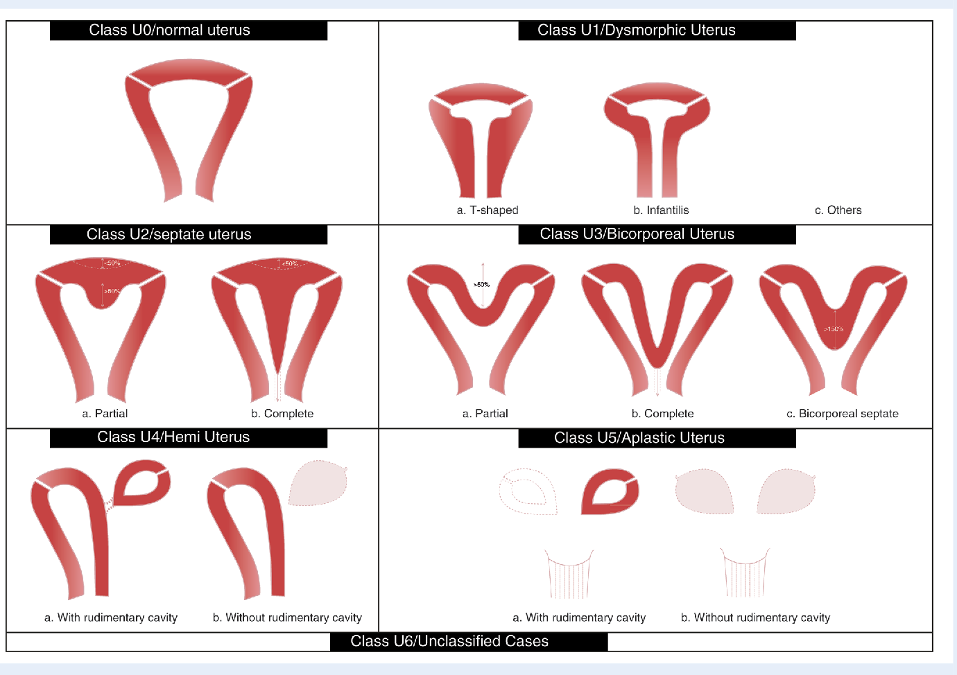
According to the ESHRE / ESGE classification, several types of anomalies exist.

The most common is the intrauterine septum (up to 35–55% of cases). It is most closely associated with miscarriage: the risk of miscarriage can reach 60–80%.

A bicornuate uterus increases the risk of premature birth.
A unicornuate uterus is associated with a high risk of pregnancy complications. Also, with these specific uterine development features, we see a high predisposition to a thin endometrium (endometrial hypoplasia), which specifically complicates embryo attachment.

Rare conditions, such as uterine aplasia, occur in approximately 1 in 4500–5000 women.

Let’s look at specific features of the uterine body structure in more detail.

Changes in uterine structure – how the uterus is formed and why sometimes it's 'not according to plan'.

In 2013, the European Society of Human Reproduction and Embryology and the European Society of Gynaecological Endoscopy (ESHRE/ESGE) proposed a new classification of female genital tract anomalies, based on the anatomy of the organ with clearly defined anatomical categories.
Class U0 – includes normal uterine morphology. A normal (typical) uterus has a pear shape externally and a triangular cavity internally; the base of the triangle faces upwards, and there may be a slight indentation that should not exceed 50% of the uterine wall thickness.
Class U1 – cases with normal uterine contours but a modified shape of the uterine cavity, excluding septa. This class includes the T-shaped uterus, characterized by a T-shaped cavity, as well as the uterus that is anatomically reduced in size.
Class U2 – the uterus has normal external contours, but a longitudinal intrauterine septum is present. This structural variant arises from incomplete resorption of the midline septum during fetal development. The septum can be either partial (small size) or complete (extending across the entire plane of the uterine body).
The intrauterine septum is the most common among atypical uterine structures. Hysteroscopic metroplasty is currently recognized as the “gold standard” for treating women with a uterine septum. Most publications indicate that indications for such surgery are spontaneous miscarriages, premature births, and conception problems when endocrine, infectious, and other possible causes are excluded. That is, if this is an incidental finding during an examination without complaints, this condition does not require any surgical treatment.
Class U3 – such a uterus looks like a “heart” both externally and internally. On ultrasound images, we see a specific indentation in the middle of the external and internal contours of the uterus, forming two semi-cavities. A bicornuate uterus is formed between the 10th and 14th week of intrauterine development; if incomplete fusion of the Müllerian ducts occurs during embryogenesis, various variants of the bicornuate uterus are formed. A bicornuate uterus rarely requires surgical reconstruction.
Class U4 – a unicornuate uterus has an elongated shape and is turned to the left or right side; the cavity loses its triangular shape and appears elongated. A unicornuate uterus occurs in situations where only one paramesonephric duct forms, while the other does not develop at all or is underdeveloped. Such a uterus may have a contralateral incompletely formed horn, which may or may not be connected to the main uterine cavity. Interestingly, in rare cases, if there is a rudimentary horn connected to the uterine cavity, an embryo can implant in it — this is a dangerous condition requiring surgical treatment. Often, these women exhibit changes in kidney structure. This is a high-risk type of uterine structure, but with proper planning and monitoring, most women can give birth to a child.
Class U5 – uterine aplasia, meaning its absence (Mayer-Rokitansky-Küster-Hauser (MRKH) syndrome). A form where a non-functioning horn exists is also distinguished. It occurs because the Müllerian ducts did not form at all or formed incompletely. This type of development is the most serious, as, unfortunately, such patients cannot conceive or carry a child. They have complete indications for gestational (surrogacy) motherhood.

What are the symptoms of uterine structure anomalies?

Clinical manifestations of uterine development disorders depend on the type of structure and the degree of changes in the internal genital organs.

Symptoms may include:

  • Absence of menstruation
  • Irregular or painful menstruation
  • Heavy or prolonged menstruation
  • Pelvic pain that may come and go
  • Discomfort or pain during sexual intercourse
  • Recurrent pregnancy loss or miscarriages
  • Infertility or difficulty with pregnancy
  • Premature birth or premature rupture of membranes
  • Abnormal fetal position during pregnancy
  • Symptoms related to urination if there are also kidney problems

Why many women are unaware of this

In most cases, there are no symptoms. Menstruation may be regular, and overall health may be normal. It is only during pregnancy planning or after a loss that the need for deeper diagnosis arises.

Diagnosis of uterine anomalies: which methods actually work

Today, the most informative method is 3D ultrasound. Since ultrasound is routine, non-invasive, and easily accessible, it can be considered the “gold standard” of diagnosis. Its accuracy reaches 95%, which significantly exceeds standard ultrasound. In complex cases, MRI or hysteroscopy is used. Key point: the correct diagnosis determines the correct tactics.

Interesting fact: the link to the kidneys

Up to 30–40% of women with uterine anomalies also have features in kidney structure. This is explained by the fact that these organs are formed simultaneously during embryonic development.
This is why examination of the urinary system is mandatory.

What this means for the possibility of getting pregnant

The presence of an anomaly does not mean that pregnancy is impossible. But it can affect:

embryo implantation,
the course of pregnancy,
the risk of miscarriages or premature births.

And that’s why it’s important not just to “know,” but to understand how this affects your specific situation.

Conclusion

Uterine anomalies are not a verdict or a reason to lose hope for motherhood. Most are structural features that do not affect daily life and its quality. An individual approach, timely monitoring, and
trust in your doctor significantly increase the chances of a successful pregnancy. Every story is unique, and for most women, a happy ending is possible. The main thing is not to be left alone with your fears and questions. Together, we can make the path to your motherhood safe and confident.

Frequently Asked Questions (FAQ)

Can I get pregnant with a uterine anomaly?

Yes, in most cases, pregnancy is possible but requires the right approach.

What is the most common anomaly?

Intrauterine septum — up to 50% of cases.

Is surgery always necessary?

No, only in the presence of clinical indications.

Do I need to check my kidneys?

Yes, up to 30–40% of cases are accompanied by anomalies of the urinary system.

List of Sources Used:

1. The ESHRE/ESGE consensus on the classification of female genital tract congenital anomalies. Grimbizis GF, Gordts S, Di Spiezio Sardo A, Brucker S, et al. Human Reproduction, Vol.28, No.8 pp. 2032– 2044, 2013 Advanced Access publication on June 14, 2013 doi:10.1093/humrep/det098
2. https://www.mdpi.com/2077-0383/11/5/1251
3. Obstetrics and Gynecology: National Guide. Vol.4: Operative Gynecology. Edited by Zaporozhan V.M. K.: VSV “Medicine” 2014.

Read also:

Cholesterol: good and bad. Norms, lipid profile, atherogenic coefficient and when treatment is needed

What high cholesterol means, how to read a lipid profile, what the atherogenic index is. Explained by Dr. Anna Vitaliivna Sharhorodska.

Secondary infertility: why pregnancy may not occur again

Secondary infertility is a condition that can affect any couple who already have children. It is not just a medical term but a reality that many face.

Sun and pregnancy: benefits, risks and rules for safe sun exposure

Pregnancy is a period when a woman’s body works twice as hard, providing not only for its own needs but also for the development of new life.

Normal progesterone and estrogen levels for cryo-embryotransfer and their impact on pregnancy success

In the effectiveness of the IVF program, preparation for frozen embryo transfer (FET) requires not only the correct endometrial thickness...

Why may pregnancy not occur with PCOS?

Polycystic Ovary Syndrome (PCOS) is a common endocrine disorder in young women. Yes, young women, because as women age, their egg reserve decreases, and over time, the number of follicles in the ovaries tends to reduce, even for those who had a very strong reserve.